
Why Should You Consider Getting a Comprehensive Eye Exam at a Trusted Local Practice?
Understanding What a Comprehensive Eye Exam Actually Involves
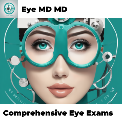
A comprehensive eye exam represents far more than simply reading letters on a chart from across a room. The experience encompasses an intricate series of tests, evaluations, and measurements designed to assess not only how clearly someone sees but also the overall health of their ocular system. Eye MD MD performs these detailed examinations with precision and care, utilizing state-of-the-art diagnostic equipment alongside time-tested clinical methodologies.
The process typically begins with a thorough case history. During this initial phase, the eye care professional asks pointed questions about current vision concerns, family history of eye disease, general health conditions, medications being taken, and lifestyle factors affecting visual function. This conversational foundation proves crucial because many eye conditions have hereditary components or connections to systemic diseases like diabetes and hypertension. Understanding this context allows practitioners to look more carefully at specific areas during the examination.
The Visual Acuity Component and Beyond
Visual acuity testing forms just one piece of the comprehensive puzzle. While most people associate eye exams with the familiar Snellen chart—that wall-mounted display with progressively smaller letters—modern comprehensive exams utilize multiple methods to evaluate clarity of vision at various distances. Digital displays, handheld charts, and specialized equipment provide measurements that painting a complete picture of refractive status.
However, detecting whether someone needs corrective lenses represents only the beginning. The examination must also evaluate:
- How well both eyes work together as a coordinated team
- The ability to focus at different distances smoothly and efficiently
- Color perception and discrimination
- Peripheral vision extent and sensitivity
- Depth perception and three-dimensional vision capability
Each component tells clinicians something vital about visual function and potential underlying problems.
Assessing Eye Pressure and Structural Integrity
Tonometry—the measurement of intraocular pressure—holds particular significance because elevated pressure can indicate glaucoma, a serious condition that frequently progresses silently without noticeable symptoms. Eye MD MD employs multiple tonometry techniques to ensure accurate readings. Variations exist across different measurement methods, and taking multiple readings provides greater confidence in the results.
The anterior segment examination allows practitioners to inspect the cornea, iris, and lens using specialized magnification equipment called a slit lamp. This powerful microscope-like device illuminates different structures of the eye, revealing inflammation, scars, cataracts, or other abnormalities invisible to the naked eye. The examination can detect early cataract formation long before vision becomes noticeably affected.
The Dilated Eye Examination Explained
Dilating the pupils through special eye drops remains one of the most important—yet sometimes misunderstood—components of comprehensive eye exams. The dilation process allows clinicians to examine the retina and optic nerve at the back of the eye, structures impossible to visualize clearly otherwise. This examination can reveal signs of:
- Diabetic retinopathy in patients with diabetes
- Age-related macular degeneration threatening central vision
- Retinal tears or detachment requiring urgent treatment
- Optic nerve damage from glaucoma
- Signs of systemic diseases affecting the eyes
Many patients feel nervous about dilation, worried about temporary blurred vision or light sensitivity. These temporary effects typically resolve within several hours, and the invaluable information gained justifies the brief inconvenience.
Why Annual Comprehensive Eye Exams Matter for Long-Term Vision Health
Regular comprehensive eye exams function as preventive health screenings comparable to dental checkups or blood pressure monitoring. Numerous eye conditions develop gradually without causing noticeable symptoms until significant damage has already occurred. By the time someone realizes something feels wrong with their vision, irreversible changes may have progressed extensively.
Consider glaucoma, often called the "silent thief of sight." This condition damages the optic nerve through elevated internal eye pressure, yet individuals experience no pain, no obvious vision changes during early stages, and no warning signs whatsoever. Only through systematic testing during comprehensive exams can eye care professionals detect this dangerous condition before it causes permanent blindness. Early detection allows intervention that halts progression and preserves remaining vision.
How Early Detection Prevents Vision Loss
Catching eye disease in its earliest stages fundamentally changes treatment outcomes. Conditions detected early typically respond better to interventions, whether through medication, lifestyle modifications, or surgical procedures. The difference between detecting a problem at the beginning versus discovering it after substantial damage represents the difference between preserving useful vision and facing vision impairment.
Age-related macular degeneration presents another compelling example. This condition primarily affects older adults, gradually destroying central vision necessary for reading, recognizing faces, and driving. Certain types progress rapidly, but medical interventions can slow progression if initiated early. Waiting until vision becomes noticeably blurred means waiting too long.
Diabetic retinopathy similarly demonstrates the critical importance of regular screening. Diabetes damages blood vessels in the retina, eventually causing vision loss if untreated. Remarkably, people can have diabetic retinopathy at various stages without realizing it. Only comprehensive eye exams with dilated pupil examination can reveal these changes. Starting treatment promptly prevents progression to vision-threatening stages.
The Role of Monitoring Existing Eye Conditions
Patients with diagnosed eye conditions or those at higher risk for developing problems benefit especially from regular comprehensive exams. Monitoring changes over time provides clinicians with objective data about disease progression or stability. This information guides treatment decisions—whether current approaches work adequately or require adjustment.
Specific Eye Conditions Detected During Comprehensive Exams
Several potentially serious eye conditions might go undetected without proper screening. Comprehensive eye exams serve as the primary detection tool for numerous problems that could otherwise lead to vision loss or indicate more serious health issues.
Cataracts and Their Progressive Nature
Cataracts develop when the eye's lens gradually becomes cloudy, scattering light and reducing vision clarity. This process typically occurs slowly over years, particularly as people age. Early cataracts might cause only minor vision changes—slightly dimmer vision, difficulty with night driving, or colors appearing less vibrant. These subtle changes often develop so gradually that people adapt without realizing change has occurred.
During comprehensive exams, Eye MD MD practitioners can detect cataracts at remarkably early stages using slit lamp examination. Identifying cataracts before they significantly impact vision quality allows for informed decision-making about when to pursue surgical intervention. Some people live perfectly well with mild cataracts for years, while others find them troublesome enough to warrant earlier treatment.
Presbyopia and Age-Related Vision Changes
After approximately age forty, virtually everyone experiences presbyopia—a gradual loss of the eye's ability to focus on near objects. This natural aging process occurs because the lens loses elasticity over time. The eye must work harder to achieve focus at close distances, leading to eye strain, headaches, and difficulty reading without holding materials farther away.
Many people don't realize presbyopia represents a normal developmental process rather than a disease or problem requiring concern. Comprehensive eye exams can distinguish presbyopia from other focusing problems and determine appropriate corrective solutions. Options range from simple reading glasses to multifocal lenses providing clear vision at multiple distances.
Refractive Errors: Myopia, Hyperopia, and Astigmatism
Refractive errors—myopia (nearsightedness), hyperopia (farsightedness), and astigmatism (irregular corneal shape)—represent the most common vision problems. These conditions develop when the eye's shape or corneal curvature prevents light from focusing precisely on the retina. While not diseases in the traditional sense, uncorrected refractive errors cause blurred vision, eye strain, and reduced quality of life.
Comprehensive exams include precise measurement of refractive error using both automated equipment and manual testing. Eye MD MD practitioners perform these measurements with careful attention to detail, ensuring the most accurate possible correction. Proper refraction makes an enormous difference in daily visual comfort and performance, whether at work, school, or leisure activities.
Dry Eye Syndrome and Surface Problems
The eye's surface requires careful balance of moisture, oil production, and tear film stability to maintain comfort and clear vision. Dry eye syndrome develops when tears don't adequately lubricate the surface, causing irritation, redness, and ironically, excessive tearing as the eye compensates for inadequate lubrication.
Environmental factors, aging, medications, and systemic conditions all contribute to dry eye development. During comprehensive exams, practitioners evaluate tear production and quality, assess lid function, and examine the corneal surface for damage. Early identification allows for treatment approaches that dramatically improve comfort and prevent progression to corneal damage.
The Technology and Equipment Behind Modern Comprehensive Eye Exams
Today's comprehensive eye exams leverage sophisticated diagnostic technology alongside classical clinical examination techniques. Eye MD MD invests in contemporary equipment that provides precise measurements and detailed imaging unavailable just years ago.
Advanced Imaging and Diagnostic Equipment
Optical coherence tomography (OCT) represents a revolutionary technology enabling practitioners to visualize the retina's internal layers with astonishing detail. This non-contact
imaging technique creates cross-sectional images of retinal tissue, revealing subtle changes that might indicate macular degeneration, diabetic retinopathy, or other retinal conditions. The technology works through light waves rather than radiation, making it completely safe for frequent use even in sensitive populations like children and pregnant individuals.
Fundus photography provides detailed color images of the optic nerve and retina. These photographs serve as permanent records allowing clinicians to compare images over time, tracking whether conditions remain stable or show progression. The comparison capability proves invaluable for monitoring glaucoma, diabetic retinopathy, and other progressive conditions.
Automated refraction equipment measures refractive error with precision, providing baseline information that practitioners then refine through manual testing. This combination of automated and subjective measurement ensures accuracy while reducing exam time.
Why Professional Expertise Remains Essential
While diagnostic equipment provides invaluable information, interpreting results and integrating findings into a comprehensive clinical picture requires extensive training and experience. An eye care professional synthesizes information from multiple tests, considers the patient's history and symptoms, and draws conclusions about what findings mean for that individual's specific situation.
Equipment can measure intraocular pressure, but only a trained clinician determines whether that specific pressure level poses risk for glaucoma in a particular patient. Imaging can show retinal changes, but distinguishing between normal variations and pathological findings requires expertise. Comprehensive eye exams represent a partnership between sophisticated technology and skilled clinical judgment.
Understanding Your Risk Factors and Personalized Screening
Not everyone requires identical screening approaches or examination frequencies. Comprehensive eye exams should incorporate awareness of individual risk factors that make certain conditions more likely or more serious if missed.
Age-Related Risk Considerations
Aging increases risk for numerous eye conditions including glaucoma, age-related macular degeneration, cataracts, and diabetic retinopathy. The American Academy of Ophthalmology recommends that adults with no eye disease and no symptoms receive comprehensive exams every five to ten years before age forty, then every two to four years between ages forty and fifty-four, every one to three years between ages fifty-five and sixty-four, and annually after age sixty-five.
However, these represent general guidelines. Individuals with specific risk factors may require more frequent screening. During comprehensive exams, Eye MD MD practitioners discuss appropriate screening intervals based on individual circumstances.
Family History and Genetic Predisposition
Certain eye conditions run strongly in families. Glaucoma, for instance, occurs more frequently in people with affected relatives. Age-related macular degeneration similarly shows hereditary patterns. When family members have experienced certain eye conditions, individuals deserve more aggressive screening and earlier baseline examinations.
Discussing family eye health history during comprehensive exams allows practitioners to adjust their evaluation focus appropriately. If a parent developed glaucoma, that information prompts more careful pressure measurement and optic nerve assessment in their adult children.
Systemic Disease and Medication Connections
Diabetes, hypertension, and autoimmune conditions all affect eye health. Diabetes particularly threatens vision through diabetic retinopathy, while high blood pressure can damage retinal blood vessels. Some medications cause side effects affecting vision or eye structures.
Comprehensive eye exams should always include detailed discussion of general health conditions and medications. This information guides which tests require particular attention and helps identify potential medication side effects affecting vision.
Occupational and Lifestyle Risk Factors
Certain occupations expose eyes to unusual stress or hazardous materials. People working with computers experience digital eye strain from prolonged screen time. Those working outdoors face increased ultraviolet exposure. Athletes and those in contact sports need evaluation for protective eyewear requirements.
Lifestyle factors like smoking accelerate age-related macular degeneration and cataract development. Alcohol consumption affects vision and eye health. Discussing these factors allows eye care professionals to provide personalized counseling about lifestyle modifications supporting long-term vision health.
The Connection Between Eye Health and Overall Wellness
The eyes provide windows into systemic health, revealing signs of diseases affecting the entire body. Comprehensive eye exams sometimes detect conditions that patients haven't yet realized they possess, leading to diagnoses requiring medical attention beyond the eye care arena.
Detecting Systemic Disease Through Eye Examination
High blood pressure damages retinal blood vessels, causing characteristic changes visible during eye examination. Diabetes damages these same vessels plus the retina itself. Autoimmune conditions like rheumatoid arthritis and lupus can cause ocular inflammation. High cholesterol deposits fatty materials in retinal vessels.
Eye practitioners trained to recognize these systemic manifestations sometimes identify problems patients should discuss with their primary care physicians. A comprehensive eye exam occasionally becomes the catalyst leading to important health discoveries.
Neurological Conditions and Vision Changes
Certain neurological disorders affect the optic nerve or vision pathways in the brain, causing vision changes that might otherwise seem mysterious. Multiple sclerosis can cause optic neuritis—inflammation of the optic nerve causing vision loss and pain with eye movement. Brain tumors can compress vision pathways, causing peripheral vision loss or other visual field defects.
Comprehensive eye exams include testing for these possibilities through careful assessment of eye movements, pupil responses, and visual fields. Identifying these findings prompts appropriate referrals for neurological evaluation.
Cardiovascular Health Indicators
Retinal blood vessel changes often mirror what's happening in blood vessels throughout the body. Examining retinal vessels provides information about cardiovascular health, atherosclerosis progression, and stroke risk. This observation holds particular value because the retina represents one of the few places where blood vessels can be directly visualized non-invasively.
Addressing Common Misconceptions About Comprehensive Eye Exams
Several misconceptions deter people from pursuing comprehensive eye exams, despite their clear importance for vision health. Clarifying these misunderstandings helps people recognize the genuine value comprehensive exams provide.
The "I See Fine" Misconception
Many people believe they don't need eye exams if their vision feels adequate. This assumption misses the fundamental point that numerous serious eye conditions progress without causing noticeable vision changes until advanced stages. Someone experiencing perfect vision today could harbor glaucoma, early macular degeneration, or diabetic retinopathy detected only through systematic testing.
Comprehensive exams serve a screening function comparable to colonoscopies or mammograms—identifying disease before symptoms appear. Waiting for symptoms to develop means waiting too long for several sight-threatening conditions.
Cost Concerns and Health Insurance Coverage
Some people avoid comprehensive exams due to cost concerns, assuming they cannot afford the examination. Many health insurance plans cover comprehensive eye exams, particularly for preventive care. Even without insurance, Eye MD MD offers transparent pricing and discussion of examination costs during scheduling.
Viewing comprehensive exams as investments in long-term vision health provides useful perspective. The cost of early detection and treatment pales in comparison to the expense and difficulty of managing advanced eye disease or vision loss.
The "My Primary Care Doctor Checks My Eyes" Assumption
Primary care physicians provide important general health screening, but their eye examinations differ substantially from comprehensive eye exams performed by optometrists or ophthalmologists. Primary care providers conduct brief eye assessments checking basic visual function, but they lack specialized equipment and training for detailed ocular evaluation. Comprehensive eye exams represent a distinctly different service providing information primary care doctors cannot obtain during general physical examinations.
Misconceptions About Dilated Pupils
Some people avoid comprehensive exams due to concerns about pupil dilation. While temporary blurred vision and light sensitivity do occur, these effects resolve completely within several hours. The information gained through dilated examination vastly outweighs the temporary inconvenience. For individuals unable to tolerate dilation due to work schedules or other commitments, scheduling exams strategically—perhaps before days off—solves the practical concern.
What to Expect During Your Visit to Eye MD MD
Understanding what happens during a comprehensive eye exam helps people prepare mentally and practically for their appointment.
Pre-Examination Preparation and Initial Intake
Arriving several minutes early allows time for check-in procedures and completion of patient history forms. These forms gather detailed information about current eye concerns, previous eye procedures or injuries, family eye disease history, and general health conditions. Bringing current medications or their list helps practitioners assess whether any drugs might affect vision or eye health.
The Comprehensive Testing Sequence
The examination typically begins with automated refraction measurements, followed by manual refinement of these measurements through subjective testing—the "better one or two" comparisons. Visual acuity testing at multiple distances follows, then assessment of eye coordination and focusing ability.
Intraocular pressure measurement (tonometry) comes next, using either contact methods or non-contact air puff techniques. Practitioners then examine external eye structures and the anterior segment using the slit lamp microscope. Visual field testing may be performed, particularly for patients at glaucoma risk or those with existing glaucoma.
Finally, pupils are dilated for posterior segment examination, allowing practitioners to evaluate the retina and optic nerve directly. Depending on findings and individual circumstances, additional tests like OCT imaging or fundus photography might be performed.